


")


Scientists have just debuted a new way to see how cells organize themselves, shedding modern light on the building blocks of life.
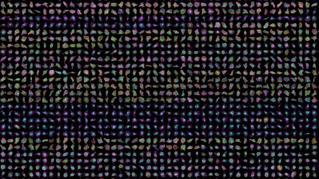
In a new database of 200,000 images, scientists captured details about the rich variation in their shapes—even among genetically identical cells grown under the same conditions.
Published in the journal Nature this week, the research is the culmination of all the work the Allen Institute for Cell Science has been doing since it was launched 8 years ago.
This milestone in cell biology—akin to discovering “design principles” of the cell—unlocks the potential to find new treatments for diseases where cells malfunction—and the methods and findings are generalizable to virtually any cell.
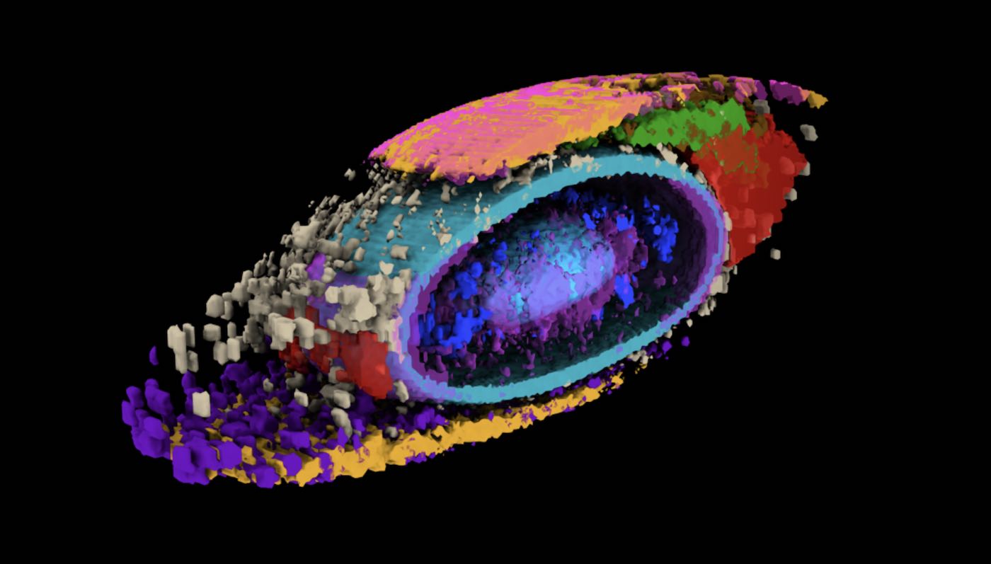
For the study, researchers created a new method of analyzing human cells that produces a new type of information beyond genomics: computationally derived, 3D spatial organization and morphology—essentially, a cell’s shape and how its internal components are organized inside in three dimensions.
After they applied numbers and mathematical principles to cell organization, they uncovered the endless variation in cell shapes.
Using computational analyses, researchers developed what they call a “shape space” that describes external shape. This includes things like volume, elongation, and the “pear-ness” or “bean-ness” of its shape.
ANOTHER DISCOVERY: Regenerative Medicine Breakthrough: Cellular ‘Glue’ Heals Wounds, Potentially Regrows Nerves and Tissue
The new data type will allow researchers to uncover the foundational principles of shape and internal organization. Understanding how cells organize their parts under healthy conditions—and the range of variability within “normal”—is key to understanding what goes wrong in disease.
Some early organizing principles we’ve discovered include:
- Cells organize their internal structures in similar ways despite a wide variation in shape, demonstrating a “robustness of organelle location within a cell”
- Position matters: cells at the edges of colonies seemed to have a specific shape and arrangement of organelles inside. These cells also have different protein expressions.
“Part of what makes cell biology seem intractable is the fact that every cell looks different, even when they are the same type of cell,” said Wallace Marshall, a Professor of Biochemistry and Biophysics at the University of California in San Francisco, and member of the Allen Institute’s Advisory Board.
RELATED: In a World First, Scientists Use Artificial DNA to Kill Cancer Cells
“This same variability that has long plagued the field is, in fact, an opportunity to study the rules by which a cell is put together (and) I expect that many others will adopt the same methodology.”
This paper also lays the groundwork for understanding a cell’s operating system, especially how three important factors relate—organization, behavior, molecular identity.
“We built all of this from scratch, including the metrics to measure and compare different aspects of how cells are organized,” said Ru Gunawardane, Ph.D., the Executive Director of the Allen Institute for Cell Science.
CHECK OUT: The Achilles Heel for Glioblastoma Cancer Discovered—a Rogue Protein that Turns Natural Defenses Off
“What I’m truly excited about is how we and others in the community can now build on this and ask questions about cell biology that we could never ask before.”
SHARE the Breakthrough With Science Lovers on Social Media…