


")


")


Alzheimer’s has been reversed in mice after scientists at the University of Illinois-Chicago boosted the formation of new brain cells, a breakthrough that could lead to new treatments.
Their gene therapy fueled new neurons in the hippocampus—a region in the brain vital for learning and remembering where you put your car keys.
Experiments have shown this growth process is impaired—particularly in the hippocampus—in patients and mice with mutations linked to Alzheimer’s.
The team found that the increasing production of neurons transformed the lab rodent’s defects, as the new neurons were incorporated into memory circuits, restoring normal function.
Brain cells send electric signals. We keep producing them throughout our lives, with help from our neural stem cells. But numbers tail off as we age—and fall dramatically in Alzheimer’s.
“This is the first time that there is evidence that neurogenesis plays an active role in Alzheimer’s disease pathology,” said lead author Professor Orly Lazarov. “Our discovery really opens up a huge opportunity for new therapies to develop in the field that are based on the enhancement of neurogenesis.”
In the study, published in the Journal of Experimental Medicine, stem cell survival was enhanced by deleting a gene called Bax, which is related to cell death, in the neurons of the mice.
This led to the maturation of more neurons. Follow-up testing showed that the altered mice performed better in spatial recognition and contextual memory tasks, which included finding their way around a maze.
POPULAR: Large Study Suggests Doing Chores May Be Linked to a 21% Reduced Risk For Alzheimer’s Disease
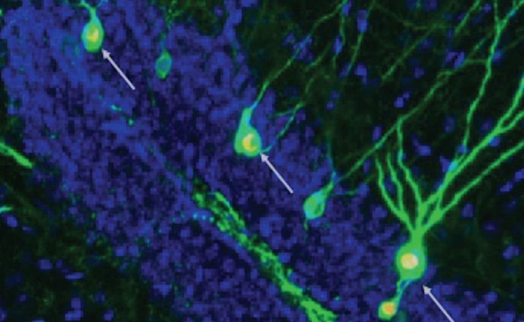
Scans of healthy mice showed the circuits involved in storing memories include many newly-formed neurons alongside older ones.
Fluorescent labels were added to the neurons, which lit them up as they were activated during memory acquisition and retrieval. Typical Alzheimer’s mice show a lack of new neurons.
Further analyses revealed there was also a rise in the number of tiny protrusions called dendritic spines. They connect neurons and are critical for memory formation.
Integration of newly formed brain cells was restored when neurogenesis was increased. When the researchers specifically inactivated the new neurons, mice with dementia lost any improvement in memory, which confirmed the results.
CHECK OUT: Eating Oily Fish Like Salmon May Cut Risk of Developing Alzheimer’s by Nearly 50%
Their study is the first to show impairments in hippocampal neurogenesis play a role in the memory deficits associated with Alzheimer’s by decreasing the availability of immature neurons for memory formation. Before the neurogenesis-based therapy can be tested in humans, the team’s findings in mice must be confirmed by other trials.
Lazarov said this work could lead to a whole new spectrum of medications that could restore memory in patients—and it offers hope to the Alzheimer’s community because current drugs target just the symptoms, not the cause.
MORE: Sunshine Could Ward Off Dementia and Strokes—First-Ever Direct Link to Vitamin D Found
DON’T FORGET to Share the Hope With Friends on Social Media…
Good news indeed. “Threshold moments” with the fridge door open help keep *me* interested in the topic, symptomatic or not.
😀
LOL. True dat!
Research indicates our memories are still located in our brains, but something is blocking our access to them. When the blocks are removed, we can access any and all of our memories… but I still like to look at photos and videos of people I’ve known and where I’ve been. 😉