














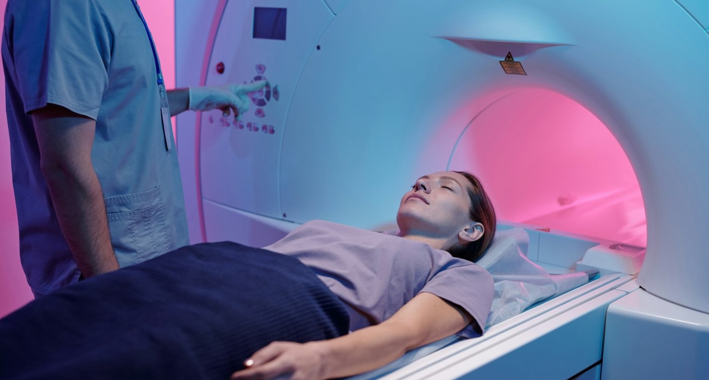
For caregivers of children living with chronic or rare lung diseases, monitoring their condition presents an uncomfortable tradeoff: Repeated scans can be valuable, but they can also expose patients to radiation.
Now, a rapidly advancing form of MRI is offering another possibility: detailed images of children’s lungs that can reveal not only their structure, but how well they’re actually working—all without exposing them to ionizing radiation.
Scientists at the University of Sheffield’s POLARIS research group have been pioneering one particularly promising version of the technology, using a specially prepared form of xenon gas.
Their work is already helping doctors identify early signs of lung disease in children with cystic fibrosis without using radiation, and the technique is now being used in patient care at Sheffield Children’s Hospital.
Lungs Are Difficult to Scan for MRIs, But There’s an Ingenious Work-Around
MRI is already widely used throughout medicine because it produces detailed images using magnetic fields and radio waves rather than ionizing radiation. But lungs have traditionally been one of MRI’s toughest challenges because they’re mostly filled with air, which produces relatively little of the conventional ‘MRI signal’, and they’re constantly moving as a patient breathes.
The Sheffield researchers found an ingenious way around that problem. Patients inhale a specially prepared form of xenon called hyperpolarized Xenon-129, which creates a strong signal that an MRI scanner can detect inside the lungs.
“Patients are asked to inhale the Xenon gas, hold their breath for a few seconds while the scanner collects the images and then breathe the gas away,” explained Professor Jim Wild, who leads the POLARIS research group.
The resulting images allow doctors to see where air is and isn’t reaching inside the lungs. Because some of the xenon also dissolves through lung tissue and into the bloodstream, researchers can gather information about one of the lungs’ most important jobs: transferring gases from the air we breathe into the blood that carries them throughout the body.
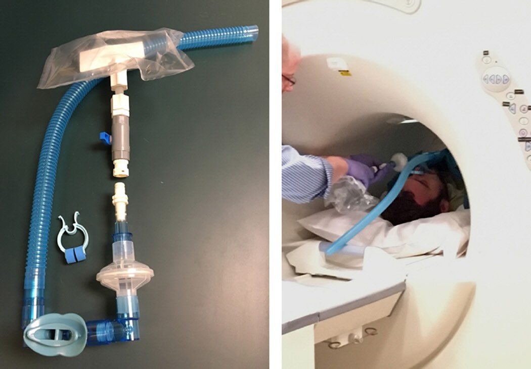
Combined with other advanced MRI techniques, doctors can potentially study ventilation, blood flow, lung structure and gas exchange without exposing the patient to ionizing radiation.
Instead of simply giving physicians a picture of the lungs, the technology can provide something closer to a map showing how the lungs are actually functioning.
That’s especially promising for children with diseases requiring years of monitoring.
BREATHING GOOD NEWS: MIT Develops AI that Predicts Lung Cancer Risk up to 6 Years in Advance, Like Finding a ‘Needle in a Haystack’
It Finds Spots That Other Tests Often Miss
Researchers have investigated pulmonary MRI in conditions including cystic fibrosis, severe asthma, interstitial lung disease, and pulmonary vascular disorders—and the Sheffield team says its xenon MRI technique has revealed lung abnormalities that routine tests did not detect.
In children with cystic fibrosis, POLARIS researchers uncovered details of ventilation difficulties that had not been picked up on previous CT scans. Sheffield Children’s Hospital is now using the MRI method to detect early functional lung disease in these patients, which the university says is the first of its kind in the UK.
The ability to look repeatedly, without adding radiation exposure, could be invaluable for a child diagnosed with such lifelong conditions.
And best of all, for the kids, the scan takes only a few minutes.
Speed is improving, too. Some newer pulmonary MRI techniques can acquire images in as little as 4 seconds—especially valuable when a patient is a child who finds it difficult to remain perfectly still or hold their breath for long periods.
Existing MRI Machines Can Be Retro-fitted for Xenon
The team is now collaborating with GE HealthCare to develop hardware and software that could make hyperpolarized xenon imaging possible on existing MRI systems. POLARIS is also supporting new clinical sites around the world with the equipment and expertise necessary to perform the scans.
Wild said the collaboration could “make the revolutionary imaging available to more patients”.
COPY THIS LINK to Share With Health Advocates On Social Media…