


")


Scientists have uncovered unprecedented insights into the formation of human hands and feet and the intricate processes that govern their development.
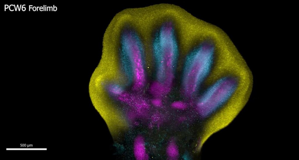
Human fingers and toes do not grow outward; instead, they form from within a larger foundational bud, as intervening cells recede to reveal the digits beneath.
After around seven weeks of cell development, an “orchestrated cell death” finally unveils the well-defined shapes of fingers or toes.
This is among many processes captured for the first time as scientists unveil a spatial cell atlas of the entire developing human limb.
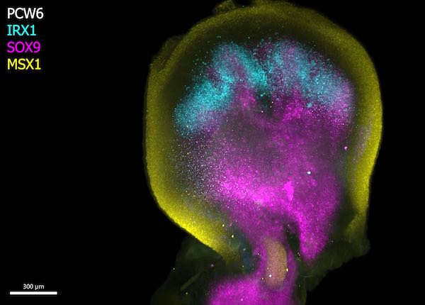
Special staining of the tissue revealed clearly how cell populations differentially arrange themselves into patterns of the forming digits.
The research could offer potential for treating muscle-related disorders or injuries, and impact the diagnosis and treatment of congenital limb syndromes.
In the study, which is part of the Human Cell Atlas initiative to map every cell type in the human body, researchers at the Wellcome Sanger Institute, Sun Yat-sen University, EMBL’s European Bioinformatics Institute and collaborators, applied cutting-edge single-cell and spatial technologies to create an atlas characterizing the cellular landscape and pinpointing the exact location of cells of the early human limb.
The atlas, published this month in Nature, provides an openly available resource that captures the intricate processes governing the limbs’ rapid development during the early stages of limb formation.
The atlas also uncovers new links between developmental cells and some congenital limb syndromes, such as short fingers and extra digits.
CHECK OUT: Woman Becomes First Human to be Fitted with Nerve and Bone Fused Bionic Limb
Limbs are known to initially emerge as undifferentiated cell pouches on the sides of the body, without a specific shape or function. But after 8 weeks of development, they are well differentiated, anatomically complex and immediately recognizable as limbs, complete with fingers and toes.
This requires a very rapid and precise orchestration of cells. Any small disturbances to this process can have a downstream effect, which is why variations in the limbs are among the most frequently reported syndromes at birth, affecting approximately one in 500 births globally.
While limb development has been extensively studied in mouse and chick models, the extent to which they mirror human situation remained unclear. However, advances in technology now enable researchers to explore the early stages of human limb formation.
In this new study, scientists from the Wellcome Sanger Institute, Sun Yat-sen University, and their collaborators analyzed tissues between 5 and 9 weeks of development. This allowed them to trace specific gene expression programs, activated at certain times and in specific areas, which shape the forming limbs.
POPULAR: Oldest Fossilized Human Footprints in North America Over 20,000 Years Old: New Study
As part of the study, researchers demonstrated that certain gene patterns have implications for how the hands and feet form, identifying certain genes, which when disrupted, are associated with specific limb syndromes like brachydactyly—short fingers—and polysyndactyly, extra fingers or toes.
The team were also able to confirm that many aspects of limb development are shared between humans and mice.
Overall, these findings not only provide an in-depth characterization of limb development in humans but also critical insights that could impact the diagnosis and treatment of congenital limb syndromes and offer potential for treating muscle-related injuries.
“Decades of studying model organisms established the basis for our understanding of vertebrate limb development,” explained Professor Hongbo Zhang, senior author of the study from Sun Yat-sen University in Guangzhou, China. “However, characterizing this in humans has been elusive until now.”
“What we reveal is a highly complex and precisely regulated process. It is like watching a sculptor at work, chiseling away at a block of marble to reveal a masterpiece. In this case, nature is the sculptor, and the result is the incredible complexity of our fingers and toes.”
AMAZING: Scientists Regrow Retina Cells to Tackle Leading Cause of Blindness Using Nanotechnology
Dr Sarah Teichmann, senior author of the study from the Wellcome Sanger Institute, and co-founder of the Human Cell Atlas, said, “For the first time, we have been able to capture the remarkable process of limb development down to single cell resolution in space and time.
“Our work in the Human Cell Atlas is deepening our understanding of how anatomically complex structures form, helping us uncover the genetic and cellular processes behind healthy human development, with many implications for research and healthcare.