


")

Brain surgery has been performed on a baby still inside the womb in order to fix potentially deadly damage to vessels and saved the infant from suffering heart failure or stroke after birth.
It was the first treated patient in a clinical trial that is underway at Boston Children’s Hospital and Brigham and Women’s Hospital, performed with oversight from the U.S. Food and Drug Administration.
Fetuses with the rare pre-natal condition known as Vein of Galen malformation (VOGM) have arteries carrying high-pressure blood that are connected to one of the main veins deep at the base of the brain. In normal fetal development, they should link to smaller capillaries, thus slowing the flow and delivering oxygen to surrounding tissue.
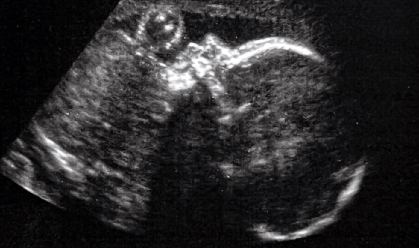
The U.S. team used ultrasound to carry out the successful procedure for a woman who was 34 weeks pregnant.
The unnamed child was delivered two days later during a normal birth after her labor was induced, due to broken membrane. The child was kept in the neonatal intensive care unit for several weeks, but mother and baby are now together at home.
“In our first treated case, we were thrilled to see that the aggressive decline usually seen after birth simply did not appear,” said lead author Professor Darren Orbach of Boston Children’s Hospital.
Repeated echocardiograms after birth displayed marked improvement in cardiac output. Scans showed normal heart and brain function.
“We are pleased to report that at six weeks, the infant is progressing remarkably well, on no medications, eating normally, gaining weight and is back home,” said Dr. Orbach in a statement.
“There are no signs of any negative effects on the brain.”
Experts described the method and outcome as “pioneering” in Stroke, the peer-reviewed journal of the American Stroke Association. The premature newborn did not require any cardiovascular support or surgery following the treatment. It had a normal neurological exam and showed no strokes, no fluid buildup, and no hemorrhage on brain MRI scans.
“This approach has the potential to mark a paradigm shift in managing Vein of Galen malformation where we repair the malformation prior to birth and head off the heart failure before it occurs, rather than trying to reverse it after birth.
“This may markedly reduce the risk of long-term brain damage, disability, or death among these infants.”
The high blood flow usually has an even more serious effect on the heart and brain after birth, putting enormous pressure on the newborn’s heart and lungs. This may lead to pulmonary hypertension, heart failure or other life-threatening conditions.
CHECK OUT: New Brain Implant Device Could Restore Function in Paralyzed Limbs
It is estimated that VOGM, the most common congenital vascular brain malformation, occurs in as many as one in every 60,000 births. It’s most often detected on a pre-natal ultrasound scan (then diagnosed by MRI) during the late second or third trimester of pregnancy.
Current standard of care is treatment after birth with embolisation, a catheter-based procedure to close off the direct artery-to-vein connections and block excess blood flow to the brain and heart. However, embolisation itself is high risk and is not always successful at reversing heart failure. Additionally, severe brain damage may have already occurred at birth, which may lead to life-long cognitive disabilities and life-threatening conditions for the infant, or even to death.
Professor Gary Satou, a fetal cardiologist at the University of California, Los Angeles, who was not involved with the study, said the intervention may be “very impactful” in a specific group of patients.
“As always, a number of these fetal cases will need to be performed in order to establish a clear pattern of improvement in both neurologic and cardiovascular outcomes. Thus, the national clinical trial will be crucial in order to achieve adequate data and, hopefully, successful outcomes.”
TEARS OF JOY: Preemie Born the Size of a Superman Doll Now Poses For Photos at Every Birthday With His Sidekick
Prof. Colin Derdeyn, a neuro-interventional radiologist at the University of Iowa, who was not involved with the study, also cautiously welcomed the breakthrough.
“The key advance here is to intervene before the physiologic events of birth can cause life-threatening heart failure. There are caveats; one successful case is not enough experience for us to conclude that the risks of this procedure are worth the benefits.
“However, the positive hemodynamic changes that they observed in utero and after birth —reduction in flow, reduction in size of the draining vein, reversal of the abnormal reversed flow in the aorta—are really encouraging.
“These are some of the most exciting and surprising aspects of this case report. This is pioneering work being done in a very careful and responsible way.”
REVEAL This Ultrasound News To Your Friends By Sharing on Social Media…